• University of California, Los Angeles
Research Cores
  • Cores
    • Animal Models & Preclinical Research
    • Biorepository & Database
    • Cellular & Molecular Analysis
    • Center for AIDS Research
    • Clinical Research
    • Data Sciences
    • Imaging
    • Materials Science
    • Microbiome Research
    • Plant Taxonomy
    • Research Coordination
  • University of California, Los Angeles

Search

Try looking up topics of interest.
Breadcrumb
  1. Home
  2. Imaging

Imaging

Cores

  • Animal Models & Preclinical Research
    • Animal Core
    • Behavioral Testing Core Facility
    • Crump Preclinical Imaging Technology Center
    • Goodman-Luskin Gnotobiotics Core
    • Mouse Physiology Lab
    • Small Animal Physiology Core
    • Translational Research Imaging Center
    • Zebrafish Core Facility
  • Biorepository & Database
    • Biorepository Core
    • Brain Tumor Translational Resource
    • Cardiac Muscle Cell Core
    • Goodman-Luskin Clinical Studies and Database Core
    • Stem Cell Bank
    • Translational Pathology Core Lab (TPCL)
  • Cellular & Molecular Analysis
    • BSCRC Flow Cytometry Core
    • Flow Cytometry Core Lab
    • IDDRC Cells, Circuits and Systems Analysis Core
    • Immune Assessment Core (IAC)
    • Immunogenetics Center (UIC)
    • Inflammatory Biology Core Lab
    • Lipidomics Lab
    • Liver Spatial Omics Core
    • Metabolomics Center
    • Mitochondrial and Metabolism Core
    • Neuroscience Genomics Core (UNGC)
    • Protein Expression Technology Core
    • Proteome Research Center
    • Sequencing Core
    • Stem Cell Engineering Center
    • Technology Center for Genomics & Bioinformatics (TCGB)
    • UCLA-DOE and Biochemistry Instrumentation Core Facility
  • Center for AIDS Research
    • CFAR Clinical Science Core
    • Centralized Lab Support Core
    • Community Engagement and Clinical Informatics Core
  • Clinical Research
    • Brain Mapping Center
    • Human Gene and Cell Therapy Facility
    • Pulmonary Function and Respiratory Physiology Lab
    • Translational Cell Therapy Lab
    • UC Fit Digital Health
  • Data Sciences
    • Biocomputing Technology Core
    • Collaboratory of the Quantitative and Computational Biosciences
    • Computing Technologies Research Lab
    • Department of Medicine Statistics Core
    • Goodman-Luskin Integrative Biostatistics and Bioinformatics Core
    • The Semel Institute Biostatistics Core
  • Imaging
    • BRI Electron Microscopy Core
    • BRI Microscopic Techniques Core
    • BSCRC Microscopy Core
    • CNSI Advanced Light Microscopy and Spectroscopy Lab
    • CNSI Electron Imaging Center for Nanosystems
    • Center for Computer Vision & Imaging Biomarkers
    • Functional Visualization Core
    • Goodman-Luskin Neuroimaging Core
    • J.D. McCullough X-ray Facility
    • Radiology Media Center
    • Translational Research Imaging Center
    • X-ray and EM Structure Determination Core
  • Materials Science
    • Analytical Phytochemical Core
    • CNSI Nano and Pico Characterization Lab
    • IGP Nuclear Magnetic Resonance (NMR) Core
    • Molecular Instrumentation Center
      • High Throughput Synthesis and Catalysis Facility
      • Magnetic Resonance
      • Mass Spectrometry Facility
      • Materials Characterization Lab
      • Microscopy Lab (MicroED & CryoEM)
    • Pasarow Mass Spectrometry Lab
  • Microbiome Research
    • Biostatistics and Bioinformatics Core
    • Goodman Luskin Human Probiotic Core
    • Goodman-Luskin Microbiome Core
    • Small Animal Imaging Shared Resource
  • Plant Taxonomy
    • Herbarium
  • Research Coordination
    • Center for Pathology Research Services (CPRS)
    • Pathology Research Portal (PRP)
    • The Center for Advanced Surgical and Interventional Technology
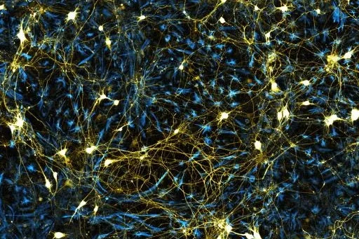
Fluorescent microscopy image of glowing neural networks on a dark background.

BSCRC Microscopy Core

Lab workers with samples

BRI Electron Microscopy Core

Lab members looking into microscopes

BRI Microscopic Techniques Core

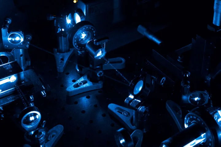
A collection of microscopes in the dark

CNSI Advanced Light Microscopy and Spectroscopy Lab

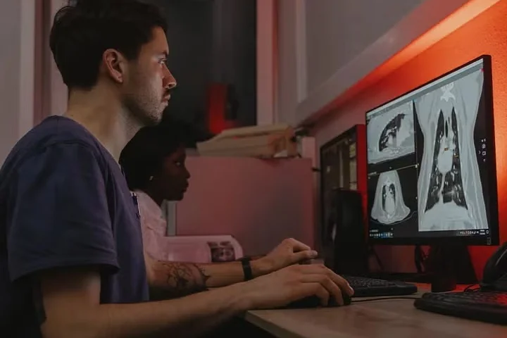
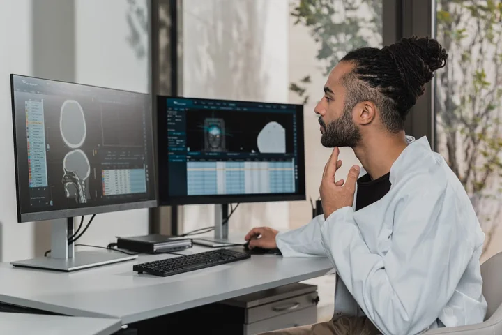
Man and woman reviewing medical scans on dual monitors.

Center for Computer Vision & Imaging Biomarkers

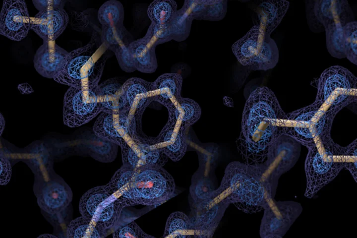
Protein structure with atomic model (yellow sticks) overlaid on a blue electron density map.

CNSI Electron Imaging Center for Nanosystems

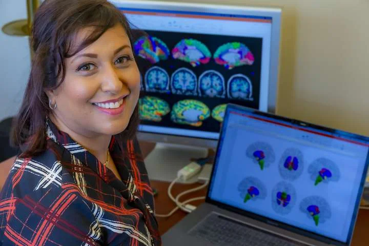
Arpana Church sitting at a desk with colorful brain imaging scans displayed on a laptop and computer screen.

Goodman-Luskin Neuroimaging Core

Scientist analyzing brain scans on dual monitors in a modern lab setting.

Functional Visualization Core

black and white photo of Professor James Douglas McCullough at his desk

J.D. McCullough X-ray Facility

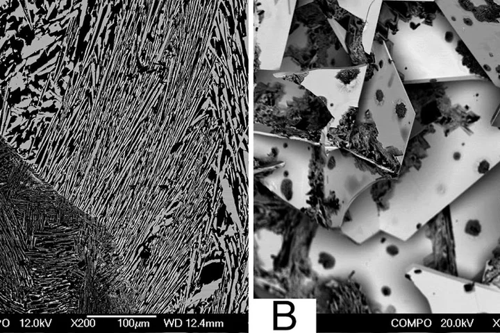
(A) SEM image of a sectioned W11B12 ingot in backscattered electron (compositional) mode indicating compositional uniformity of WB4.2 (bright) grains. (B) Backscattered electron SEM image of a fractured ingot of an arc-melted sample in the ratio W:B of 1:12. Light regions are the tungsten-containing phase.

Materials Characterization Lab

MIC lab workers at computer

Microscopy Lab (MicroED & CryoEM)

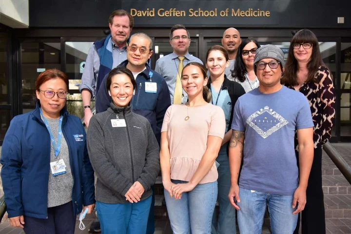
TPLC staff

Translational Pathology Core Lab (TPCL)

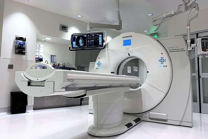
MRI system

Translational Research Imaging Center

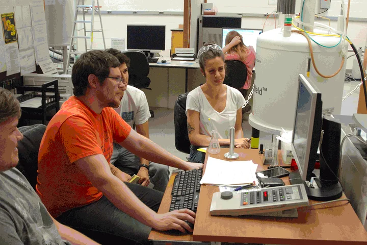
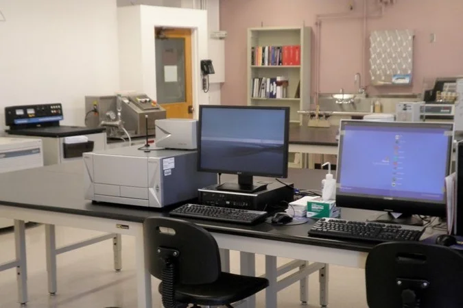
UCLA-DOE and Biochemistry Instrumentation Core facility

UCLA-DOE and Biochemistry Instrumentation Core Facility

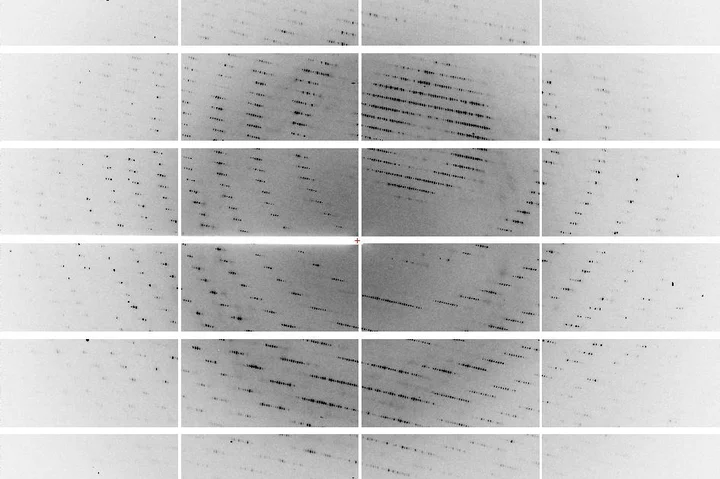
X-ray crystallography diffraction pattern showing concentric arcs of reflection spots on a segmented detector.

X-ray and EM Structure Determination Core

Home

Geffen Hall
885 Tiverton Drive
Los Angeles, CA 90095

  • Facebook
  • X-Twitter
  • Instagram
  • LinkedIn
  • YouTube
  • Connect

    • Emergency
    • UCLA Directory
    • Maps And Directions
    • Talk to Our Media Team
  • Get Involved

    • Give To DGSOM
    • Report Misconduct
    • Volunteer for UCLA Health
    • Work For UCLA Health
  • Facebook
  • X-Twitter
  • Instagram
  • LinkedIn
  • YouTube
  • Emergency
  • Accessibility
  • UCLA Privacy Policy
  • UCLA Health Privacy Notice
  • Login
© 2026 UCLA Health