Mitochondrial and Metabolism Core
Overview
The Mitochondria and Metabolism Core supports mitochondrial research for both academic and industry users. The Core specializes in mitochondrial bioenergetics and imaging, providing guidance and services focused on mitochondrial bioenergetic functions, including respirometry and ATP production, as well as imaging of mitochondrial mass, membrane potential, reactive oxygen species, and more. In addition to full service offerings and technical training, the Mitochondria and Metabolism Core is committed to providing technological resources as well as educational opportunities to further mitochondrial awareness and knowledge to the scientific community.
Leadership


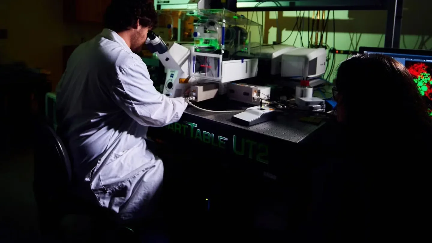
Instrumentation and Core Resources
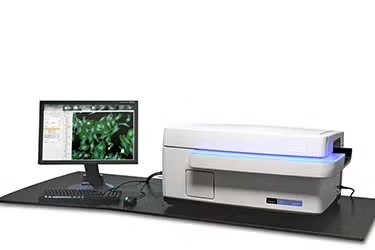
Operetta High Content Imager (PerkinElmer)
Allows investigators to conduct larger, more complex imaging experiments. Whether you are performing assay development, genome-wide siRNA, compound screens or looking at a few samples in great detail, and whether you are using 3D disease models, primary cells, stem cells, live or fixed cells, the Operetta High Content Imaging System delivers all the features you need to generate unbiased, statistically significant data to move your research forward.
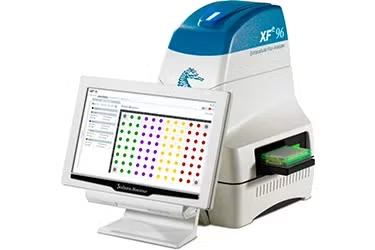
Extracellular Flux Analyzer (Seahorse/Agilent)
The Seahorse Analyzers come in a variety of models designed for different sample capacities, well formats, and cost efficiency. The Seahorse XFe96 Analyzer (2X) offers the highest capacity for XF assays with the lowest per-sample cost, allowing for large experimental groups per assay. It can also analyze individual spheroids with the Seahorse XFe96 Spheroid Microplate. The Seahorse XFe24 Analyzer (1X) uses a 24-well plate format for smaller-scale experiments. The Seahorse XF Pro (1X) features advanced software that simplifies assay setup and data interpretation, making it ideal for more complex cell types and experimental conditions, while ensuring accurate and reliable metabolic measurements. For studies with limited biological material, the XF HS Mini Analyzer (1X), with its 8-well plate format, offers high sensitivity and precision.
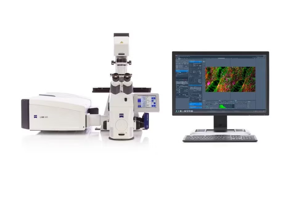
Zeiss LSM880 with Airyscan, 2-Photon and FLIM Module
The Zeiss confocal microscopy system offers high-resolution imaging of both live and fixed cells. Innovative technologies within the system enhance imaging quality and versatility, making it ideal for deep tissue visualization of cellular processes. Zeiss 2-Photon and non-descan microscopy enables imaging with low interference with samples through limiting phototoxicity.
ImageXpress High Content Confocal Screening
The ImageXpress is a high-content confocal microscope that captures high-quality images of intracellular events. This allows researchers to image and analyze mitochondrial membrane potential and morphology, ROS measurements, lysosomal function, mitophagy, and organelle interaction in an automated manner.
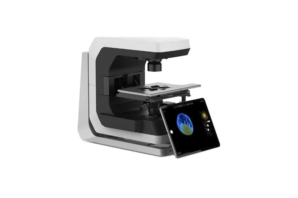
Echo Revolution
The Echo Revolution combines high-quality optics while including multi-channel image acquisition. Because of the wide range of microscopy applications, this is the perfect tool for tile-scan for fluorescence, color slides, and TC plates.
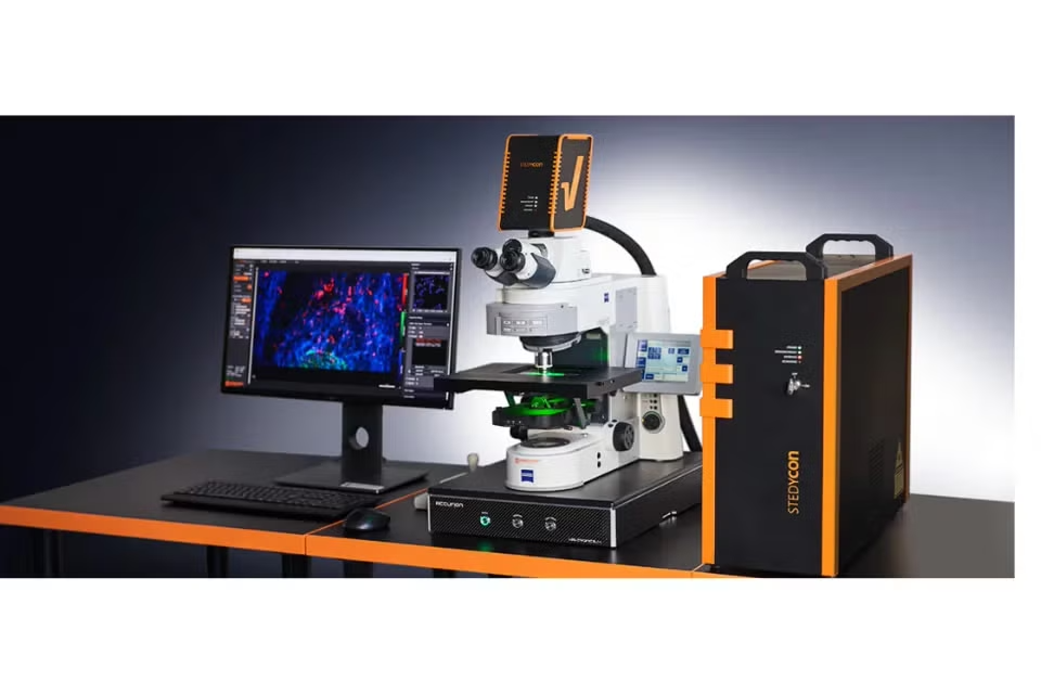
Abberior STEDycon
Allows researchers to analyze live cell imaging super-resolution of mitochondria cristae and fixed cells. STED (Stimulated Emission Depletion Super-Resolution Imaging) technology allows imaging to nanoscopic scales while providing stabilization during fast confocal imaging.
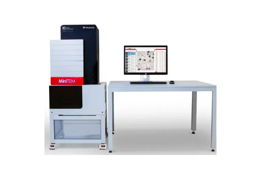
miniTEM
The miniTEM is a small but mighty instrument that performs TEM ultrastructure imaging of cells and tissues. It delivers high-resolution images revealing morphology and ultrastructure of biological samples.
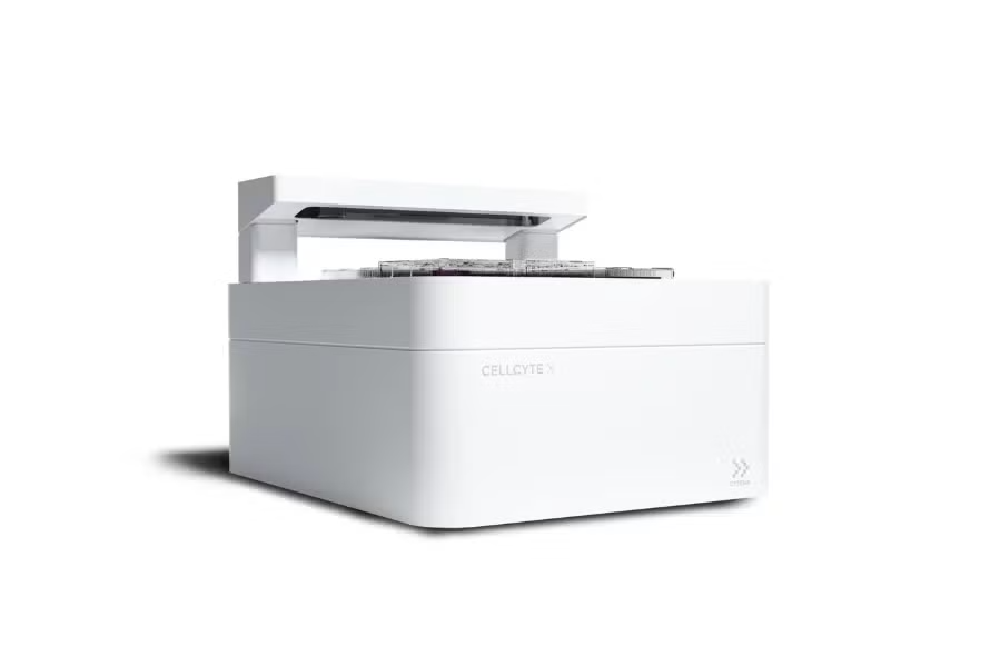
Echo Cell Cyte
The CellCyte is a powerful tool is designed for real-time, live cell observation inside the cell incubator. Researchers are able to gain insight on long-term cellular processes and kinetic behaviors in both brightfield and widefield fluorescence modes. Ideal for analyzing cell proliferation, cell death, differentiation, and target expression.
Aivia Workstation
Aivia is an advanced software with tools for two and three-dimensional image visualization and analysis. Used for trained segmentation of structures for fluorescence, widefield, and electron microscopy images analysis.