Small Animal Imaging Shared Resource
Jonsson Comprehensive Cancer Center (JCCC)
Overview
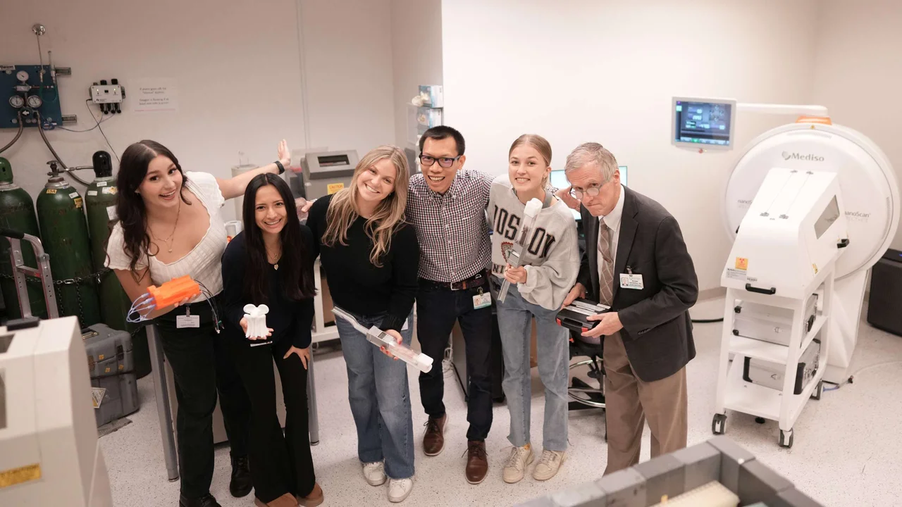
The Small Animal Imaging Shared Resource (SAISR) provides state-of-the-art in vivo imaging technologies and related diagnostic and radio-therapeutic services to JCCC and UCLA faculty, staff and students. The SAISR is a world-class, first-of-its-kind small animal imaging technology development and shared resources facility, providing JCCC investigators with technical imaging expertise and facilitating the design, execution and analysis of in vivo small animal imaging studies.
Our aims are:
- To provide expertise and support state-of-the-art small animal imaging technologies
- To provide expertise and support radiochemistry and radiolabeling production services
- To provide JCCC investigators with training in radiochemistry, preclinical imaging, and image analysis and to support the design and execution of imaging studies
The SAISR offers full-service whole body microPET, microCT, MRI, and optical imaging and complementary in vitro/ex vivo assays to measure tissue function and structure. Companion PET tracer radiochemistry and radiolabeling services are available in-house through the adjacent radiochemistry and cyclotron facility. Study design and development, training in imaging techniques, and staff assistance are offered regularly to all users.
Leadership

In Vivo Imaging Services
Nuclear imaging
- Positron emission tomography (microPET)
- Static and dynamic whole body functional imaging
- Extensive panel of radiolabeled small molecule PET tracers (readily available)
- Peptide/protein radiolabeling service and workspace
- Computed tomography (microCT)
- Whole body anatomical imaging
- Quick scan time for in vivo imaging (1-2min)
- Down to 20 µm (specimen imaging) and 100 µm (in vivo imaging) resolution
- Contrast enhancement of soft tissues such as lymph nodes, spleen, liver, heart, gastrointestinal, major blood vessels
- Cardiac gating and respiratory gating are available
- *Single photon emission computed tomography (SPECT)
Optical imaging
- Bioluminescence, Fluorescence and Cerenkov imaging
- Whole body, multi-animal imaging
- Cell localization, target trafficking, longitudinal studies
- BLI: D-luciferin substrate provided
- FLI: multiple excitation/emission filters including near-infrared
MRI imaging
- Aspect 1T preclinical MRI system (50 micron resolution) with rodent head coil and body coil
- ECG-gated cardiac MRI available
Complementary services
- Routine productions of established PET tracers
- Novel PET tracer consultation and development
- Biologics radiolabeling
- Toxicology of new PET tracers for IND submission (through UCLA DLAM)
- Imaging-based pharmacokinetics and pharmacodynamics studies
- Radiotherapeutic labeling (e.g. Lu-177, Y-90) and efficacy studies
- PET tracer whole body biodistribution and dosimetry
- PET tracer metabolite analysis
- In vivo and ex vivo biodistribution studies of imaging agents
- In vitro binding and uptake assays (e.g. radiolabeled PET tracers; fluorescent biomarkers)
- In vitro and ex vivo (tissue or whole body) autoradiography
- Quantitative image analysis and training
- Image data processing and 2D slice or 3D volume renderings
- DICOM formatting and archiving
- 3D printing of CT-scanned objects
- Rodent tail vein i.v. injection of experimental agents and cells
Equipment
- MicroPET/CT scanner (Sofie Biosciences)
- ManoScan SPECT/CT scanner (Mediso)
- CrumpCAT (microCT for live animals)
- HiCT (high-resolution microCT for sample imaging)
- M2 MRI (Aspect Imaging)
- IVIS Lumina II optical imagers (PerkinElmer)
- AMG automatic gamma counter (HiDex)
- 300SL automatic liquid scintillation counter (HiDex)
- CM3050 cryomicrotome (Leica)
- CM3600 XP cryomacrotome (Leica)
- BAS-500 phosphor imaging system (Fujfilm)
- 3D printers
- Laser cutter and engraver
Operations
- New investigators can contact the SAISR at micropet@mednet.ucla.edu to discuss potential studies, experimental design, data analysis, and for questions or advice
- After training, JCCC researchers can readily use the SAISR through secure keycard access around the clock
- Experiments are generally performed by members of the investigator’s lab after SAISR training, with expertise and assistance from the SAISR faculty and staff
- Animal housing facility: Adjacent to the Imaging Center is a barrier facility for housing animals before and after imaging with biosafety cabinets for procedures
- The Imaging Center is fully certified by UCLA’s Office of Animal Research Oversight (OARO) and Environmental Health and Safety (EH&S) for animal welfare and radiation, laboratory, biological and chemical safety procedures