Translational Pathology Core Lab (TPCL)
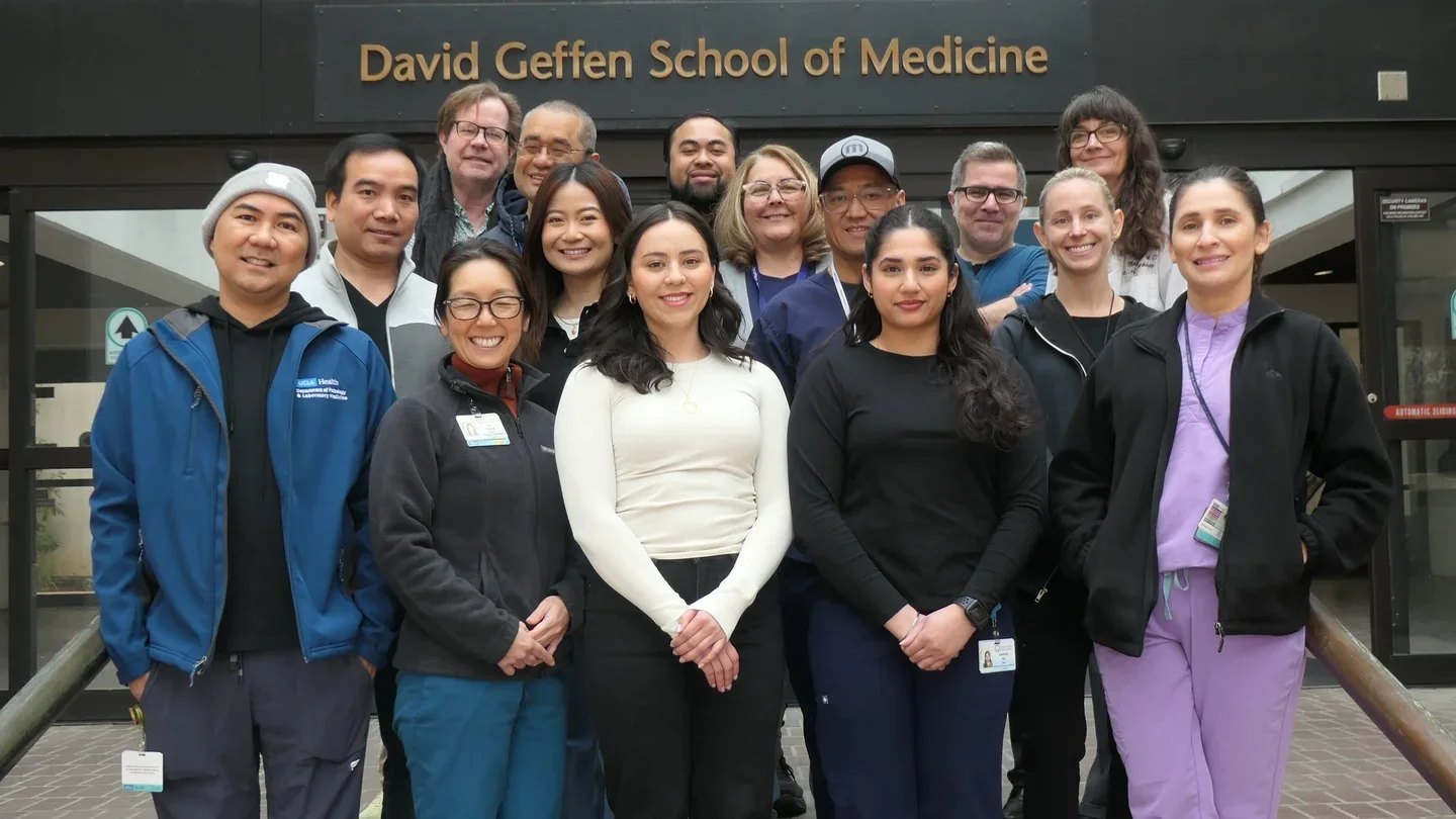
Overview
The Translational Pathology Core Laboratory (TPCL) is a CAP certified research facility in the UCLA Department of Pathology and Laboratory Medicine and a UCLA Health Jonsson Comprehensive Cancer Center Shared Facility. Since 1996, the TPCL has provided an array of pathology-related services in support of basic, translational and clinical research at UCLA. Our mission is to provide the highest quality pathology services in an efficient and cost-effective manner. The TPCL also provides expert consultative services to investigators in pathology-related study design, tissue selection, microscopic interpretation, immunohistochemistry, immunofluorescence, laser capture microdissection, digital image analysis, and IRB-related tissue request questions.
Leadership
Services
Tissue Procurement, Storage and Provision
The TPCL assists UCLA researchers with the procurement of remnant human tissues from surgical procedures. Tissue may be released fresh, snap frozen and/or formalin fixed, paraffin embedded, as well as many other specialized request. Researchers also may request tissues stored in our CAP accredited, CLIA certified biorepository, which has been procuring and storing frozen and paraffin embedded tissue since 1998. In addition, TPCL has access to the UCLA Department of Pathology clinical archive of diagnostic blocks (dating back to 1952) containing millions of tissue blocks.
The TPCL also assists with the tissue handling and processing of all of the sponsored studies at UCLA.
We also offer long term storage of biospecimens in our 24/7 temperature monitored -80 and liquid nitrogen storage facilities.
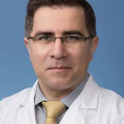
Immunohistochemical Staining (IHC)
These services are overseen by Dr. Jonathan Said, an internationally recognized expert in IHC. Services include immunostaining of established antibodies as well as optimization of new antibodies and double staining for brightfield; TUNEL assay for apoptosis is also performed. We have a large library of established antibodies available for use on human and animal tissues. Several chromogens (DAB, Fast Red, AP Blue, AP Green) and counterstains (hematoxylin, methyl-green, nuclear fast red) are available. The detection system most frequently used is the HP system using DAB with hematoxylin counterstain.
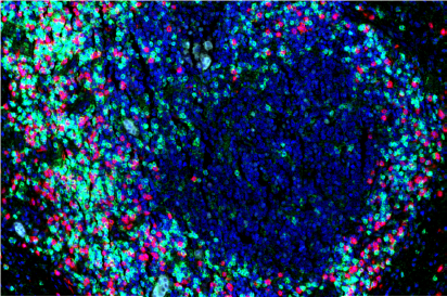
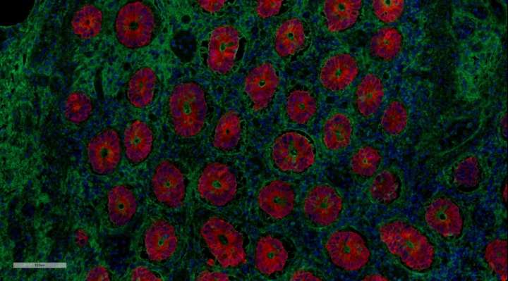
Immunoflourescent Staining (IF)
These services are overseen by Dr. Clara E. Magyar. She has over 15 years of experience in advanced microscopy and immunofluorescence staining of both human and animal tissues. Services include staining of established antibodies as well as optimization of new antibodies for immunofluorescence on formalin fixed paraffin embedded tissue sections. For multiplex staining requests, we employ the OPAL staining kit, allowing for staining of up to 4 biomarkers plus nuclear counterstain, in addition to standard immunofluorescence staining procedures.
Laser Capture Microdissection (LCM)
Laser capture microdissection (LCM) allows researchers to analyze specific cells within a larger sample. In 2010, TPCL and the California NanoSystems Institute’s Advanced Light Microscopy/Spectroscopy (ALMS) core lab jointly purchased a Leica Laser-Microdissection (LMD) 7000. This state-of-the-art LMD technology is housed in the ALMS core, and can be accessed via keycard 24/7/365 once users complete a training class. New features of this instrument include: (1) ability to capture into varied devices, including single PCR tube caps, lab-on-a-chip devices and Petri dishes; (2) a more powerful laser that allows cutting of thick (> 100 micron) sections in a single cut; (3) more precise control of the laser settings to enable collection of individual nuclei; (4) patented beam-steering technology that enables more precise and faster dissection at every magnification; and (5) gravity collection of samples that reduces contamination. Users can sign up for training at the ALMS on-line registration site. Sections must be cut onto specialized slides. The TPCL will prepare all slides for you.
Digital Imaging and Image Analysis Services
Digital imaging and image analysis services include state-of-the-art virtual microscopy (VM) and digital pathology (DP) (image analysis) services to the UCLA community. In VM, whole glass slides are converted to high resolution digital images (either brightfield or fluorescence) for easy archiving and retrieval, detailed magnification up to 40X, remote viewing via a web-based interface and preparation for publications and teaching - most of which can be performed from a single computer, using free software. DP includes performance of (or instruction in performing) quantitative digital image analysis studies. This includes both quantitative immunohistochemistry and analysis of other cellular characteristics (e.g., cell size). Dr. Clara Magyar directs the digital imaging/image analysis services for the core. The TPCL provides assistance with image acquisition and analysis, tips on sample preparation, and training on our image analysis systems. The TPCL houses several different scanners and image analysis programs (discussed in detail below). Scanners include the Applied Imaging Leica Aperio Versa high throughput scanning system (fluorescence, brightfield) and the Aperio ScanScope AT high throughput scanning system (brightfield, web enabled).Our automated digital analysis software is Definiens’ Tissue Studio.
Why Use Digital Imaging/Image Analysis?
VM/DP permits real-time discussions of histology images posted on secure websites, eliminating barriers to the exchange of pathology information with national/international collaborators/consultants, helping to reduce inter/intra-observer variability that adversely affects quantitative histopathological analysis, improving the overall efficiency of our researchers and clinicians, and eliminating delay, damage, or loss of often irreplaceable slides during shipping between UCLA faculty and extramural sites. Images can be archived, stored indefinitely, and easily retrieved - unlike glass slides, which may be lost, misfiled, damaged or fade with time.
The TPCL houses several different scanners and image analysis programs (discussed in detail below).Scanners include the Leica Aperio Versa high throughput scanning system (fluorescence and brightfield) and the Aperio ScanScope AT high throughput scanning system (brightfield, web enabled). For image analysis, we use Definiens’ Tissue Studio.
Equipment
- Leica Versa high-throughput scanning system (fluorescence, brightfield)
- Aperio AT high-throughput scanning system (brightfield, web enabled)
- Definiens’ image analysis software
- Leica laser capture microdissection LMD 700 (located in the ALMS core in CNSI)
- Revco ULT II Fischer -80 freezers
- Microm HM 550 Cryostat
- Leica RM 2135
- Microm HM 355S
- Thermo Shandon Excelsior ES tissue processor
- Embedding Machine Model HistoCenter II
- Olympus BX41 microscope.
- Leica DM1000 LED fluorescence microscope
The TCPL is a CAP accredited, CLIA certified biorepository with 24/7 temperature monitored -80 and liquid nitrogen storage capabilities.