X-ray and EM Structure Determination Core
Institute for Genomics and Proteomics (IGP)
Overview
Our Purpose
Functional Insights through 3D Structure
The X-ray and EM Structure Determination Core enables access to sophisticated equipment and technologies, offers advice, training and technical assistance in crystallization and sample preparation for CryoEM, MicroED and X-ray diffraction, data collection, processing, atomic refinement, modeling, publishing and data deposition. The core provides services of exceptionally high technical sophistication, with access to consultation and data analysis. The dedicated staff is trained to accelerate, enrich and educate structural and non-structural biologists to use imaging techniques including electron and X-ray diffraction and cryoEM. The breadth of skills and knowledge of the staff and PIs in the IGP allows us to apply any of our 3 imaging technologies as needed. The state-of-the-art resources enable the detailed 3-D analysis of biological macromolecules that play essential roles in the plant and microbial systems under investigation.
Fostering innovation
Cross-functional Synergies
The X-ray and EM Structure Determination Core conducts original research in improving crystallographic tools, continuously developing data collection techniques and processing pipelines (indexing, integration, scaling) and have added electron microscopy methods to our existing X-ray capabilities. Each structure determined using the core facility not only yields biological insights, but also expands databases used in algorithms for fold assignment, structure verification, atomic refinement, potential energy functions, and analysis of protein-protein interactions. Comprehensive databases are key to maintaining and improving these vital tools that enable reliable, high quality structure determination and prediction.
The core plays a major role in driving EM innovation and dissemination of high-resolution imaging methods; EM innovations contribute to all three projects. IGP PIs are important contributors to new EM developments, particularly the projects concerning MicroED and single-particle scaffolding for small proteins, both within the Enabling Capabilities area.
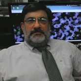
Leadership
Services
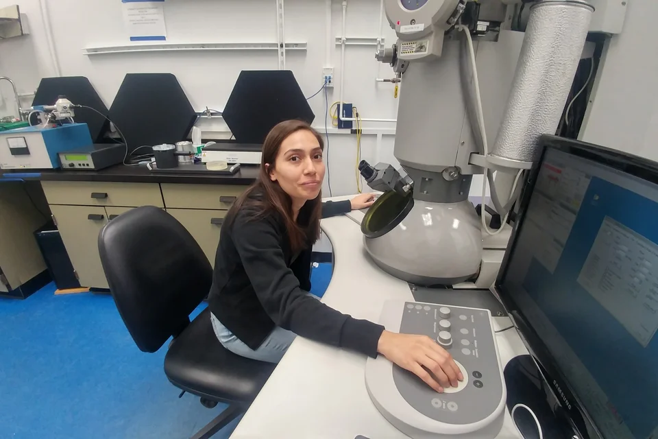
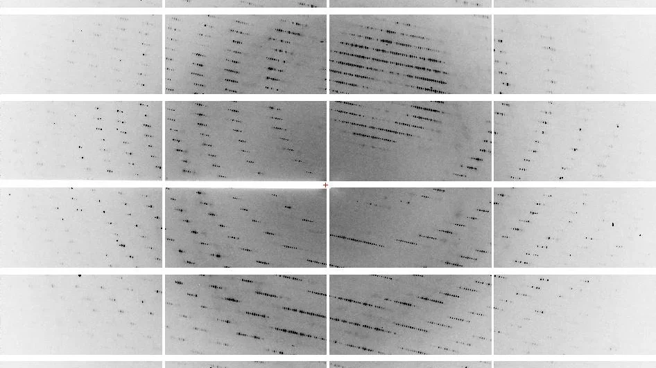
MicroED and Cryo-EM
- Facilities for high resolution electron diffraction at 300 kV on a Tecnai F30 equipped with an XF416 detector and a Talos equipped with a CetaD detector.
- Facilities to screen sample quality by negative stain imaging at 120, 200 and 300 kV
- Facilities for high resolution EM imaging including Tecnai 12, Tecnai F30 and Talos TEM microscopes and sample preparation equipment
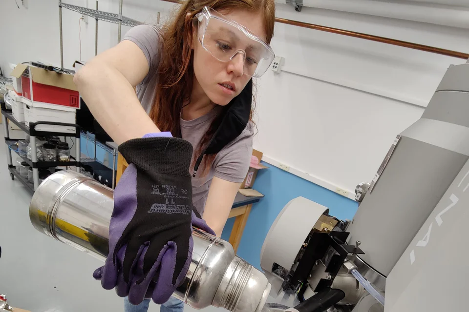
- Low dose cryo electron microscopy at 120, 200 and 300 KeV
- Computer clusters for data processing
- Experimental and computational facilities for structure analysis and refinement
- Assistance with macromolecular model building, structure refinement, and guidance in the interpretation of the resulting 3D structures
- Workshops introducing the fundamentals of Cryo-EM work and in-person training on the Electron Microscopes.
X-ray Crystallography
- Experimental and computational facilities for X-ray based structure analysis and refinement
- Acquisition of diffraction data using in-house high brilliance X-ray generators
- Acquisition of Multi-wavelength Anomalous Dispersion data using synchrotron radiation
- X-ray diffraction data processing and manipulation
- Assistance with macromolecular model building, structure refinement, and guidance in the interpretation of the resulting 3D structures
- Web-based services that include tutorials and software tools
- In-person workshops to introduce the fundamentals of single crystal X-ray diffraction and provide training on the X-ray instruments and remote data collection at the APS synchrotron.
Crystal Screening
- Consultation and technical assistance
- Evaluation of sample homogeneity and particle size distribution via Dynamic Light Scattering
- A variety of crystallization screening kits.
- Robotic setup of crystallization conditions at 4° and 20° C using TECAN, Mosquito and Echo525 liquid handlers
- UV/vis microscope to determine whether crystals are composed of protein or salt.
- Optimization of crystallization conditions
- In-person training on the instruments.
- 3D Print Prototyping
- Micro crystallography: rapid screening of micro-crystals with the Korima UV microscope, and manipulation and dissection of micro-crystals with a micro-manipulator
Facilities & Equipment
The X-ray and EM Structure Determination facility occupies 2200 square feet in total and is located in Boyer Hall. Rooms 105 and 136 are dedicated to CryoEM and MicroED, Rooms 106 and 124 are dedicated to sample preparation, characterization, crystallization screening and X-ray diffraction.
The facility incorporate emerging technologies, adding new instruments now available to IGP investigators: 1) The 300Kv Tecnai F30, 2) The 120kV Tecnai T12 TEM microscope, 3) An Acoustic Liquid Handling (Echo Labcyte) for crystallization, synthetic biology and functional screening. 4) An Octet RED96 (ForteBIO) to measure kinetics and ligand binding. 5) A Wyatt DynaPro Plate Reader to measure Dynamic and Static Light Scattering. Also we have regular access to a new 200kV Talos F200C TEM instrument that will further bolster capabilities in MicroED, CryoEM and tomography.
The facility operates an STP LabTech Mosquito nanoliter liquid handler, a Rigaku FRE+ rotating anode X-ray generator and two Rigaku HTC detectors with varimax confocal optics. Crystals are cooled at 100 K by X-tream Liquid Nitrogen cryogenic coolers. All generators are shielded by leaded glass enclosures. The facility is kept at 20 °C by two independent air conditioning systems.
The cold room also is equipped for crystal growth and mounting of heat-labile crystals. The compressors and water recirculating coolers used to remove heat from the x-ray generators are kept in the small room 116, to isolate the noise from the rest of the facility.
Pricing
To recover the cost of the supporting personnel, consumables and service contracts for the equipment, a fee will be charged to researchers.
The fee structure is designed to divide the ongoing costs among anticipated users, based on usage of the facility.
This usage covers numerous activities: x-ray data collection, machine maintenance, data collection at home, travel to synchrotrons and data collection there, expert assistance with structure determination (e.g. molecular replacement, heavy atom phasing, refinement), coordinate deposition, structure interpretation and analysis, and figure and manuscript preparation.
A point system of a unit has been developed to assign usage to participating research groups. INTERNAL X-RAY SERVICE (UCLA) will be charged the approved rate of $105.98 per unit. EXTERNAL X-RAY SERVICE ( NON-UCLA) will be charged $135 per unit. A PI should anticipate a charge of around 20 units per quarter to use every instrument, and materials available in the facility including: FRE+ X-ray generators, HTC image plate detectors, High Power Leica Microscopes with High Resolution CCD Cameras, Korima UV microscope, all Cryogenic Equipment, storage of crystals in Liquid Nitrogen Dewars, storage in the 5 degrees Crystallization Room, data collection at the synchrotron and experienced staff assistance with structure determination.
New users should discuss setting up a recharge account with the XRAY and EM STRUCTURE DETERMINATION Core Director.