CNSI Advanced Light Microscopy and Spectroscopy Lab
(ALMS)
Overview
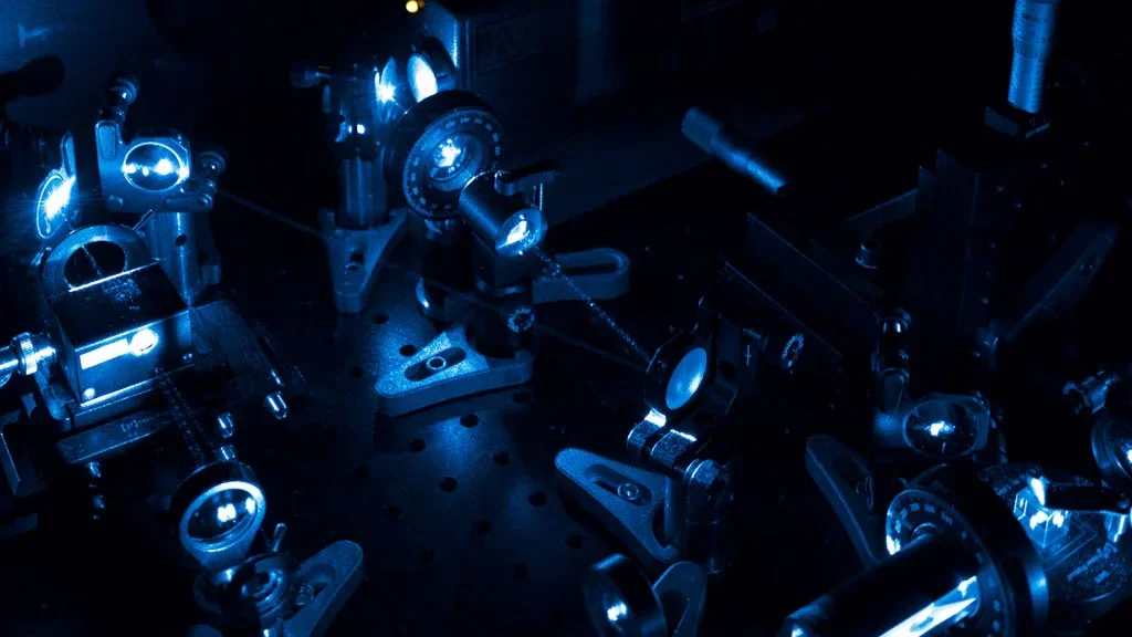
The Advanced Light Microscopy and Spectroscopy (ALMS) Lab provides comprehensive consultation, services, and training in advanced fluorescence microscopy, spectroscopy, and image analysis. Our mission is to support investigators in applying state-of-the-art microscopic and spectroscopic methods to study macromolecules, cellular dynamics, and to characterize biomaterials with nanometer precision. ALMS houses a unique collection of high-end, customized fluorescence microscopes, small-animal imaging systems, and advanced fluorescent probes, enabling fluorescence-based measurements across broad spatial (nanometers to centimeters), temporal (nanoseconds to days), and spectral (UV–NIR) ranges. Our integrated capabilities allow researchers to investigate biological processes at multiple scales, from whole in vivo animal imaging to single-molecule detection and sub-70 nm super-resolution nanoscopy, achieving exceptional spatial and temporal resolution. Beyond providing access to specialized instrumentation, we offer expert consultation and hands-on training in advanced imaging techniques and customized image analysis workflows. Our image analysis services include pixel classification, object segmentation, and tailored analytical pipelines to support complex experimental needs.
Leadership
Services
ALMS services include:
- Wide-field fluorescence microscopy
- Confocal microscopy (point-scanning and spinning disk) with tunable excitation and emission
- Two-photon laser scanning microscopy with second harmonic generation (SHG) for label-free collagen imaging
- Fluorescence correlation spectroscopy (FCS)
- Fluorescence resonance energy transfer (FRET)
- Microscopic and macroscopic fluorescence lifetime imaging microscopy (FLIM) with time-correlated single-photon counting and near-infrared detection
- Super-resolution microscopy (STED and STORM)
- Microscopic and macroscopic (small-animal) spectral unmixing
- Laser capture microdissection
- Light-sheet microscopy
- Advanced image analysis and customized workflows
- Imaging hardware customization and specialized detector integration
- Consultation and proof-of-concept studies ALMS operates as an open-access facility, serving academic investigators from local, national, and international institutions, as well as small start-up and mid-sized companies.
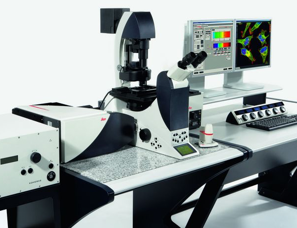
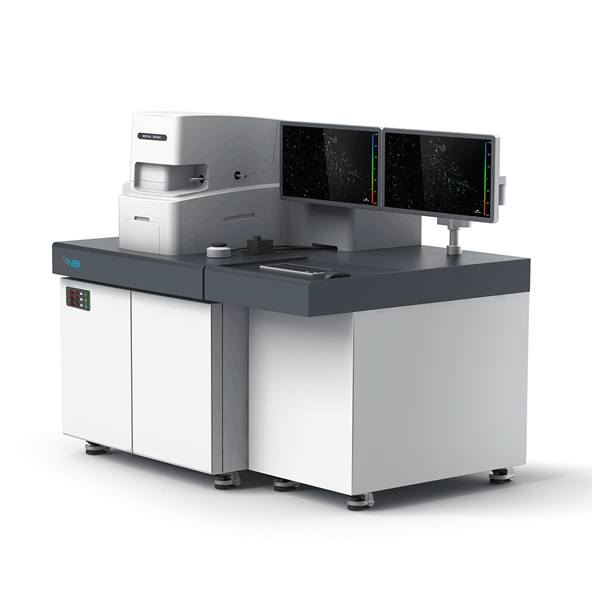
Equipment
Located on the lowest floor of the CNSI building, the laboratory features an 1,800-square-foot optical suite designed for advanced light microscopy. The space provides a tightly controlled environment with low vibration, filtered air, temperature stability (±1 °C), and light isolation to ensure optimal imaging performance.
Our laboratories offer open access to all academic investigators from local, national, and international universities, as well as to small start-up and mid-size companies.
The laboratory infrastructure includes five environmentally controlled rooms, four personal workbenches, two chemical fume hoods, one laminar flow hood, two tissue culture incubators, and two centrifuges. The facility is supported by three PhD-level technical staff. Approximately 20 workstations are available for image acquisition and analysis, with access to centralized computing resources for data transfer, storage, and printing.
Available bioimaging software includes Leica LAS X for confocal microscopy, Aivia 16 for AI-assisted image analysis and visualization, and specialized software packages for fluorescence correlation spectroscopy and lifetime analysis.
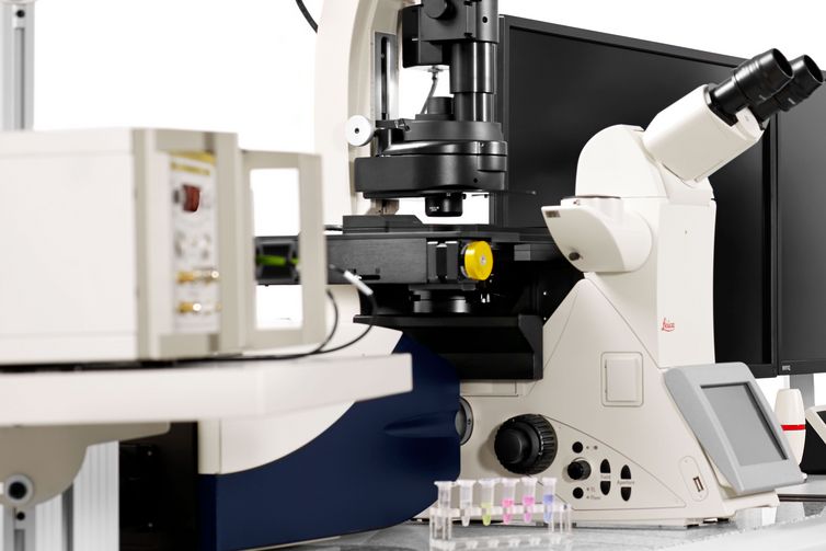
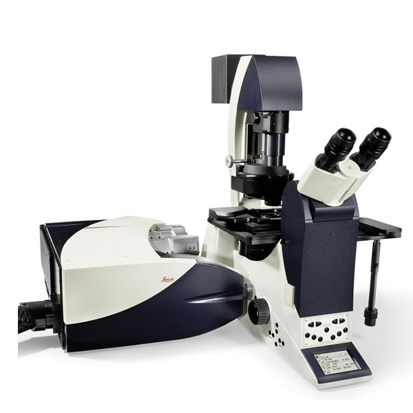
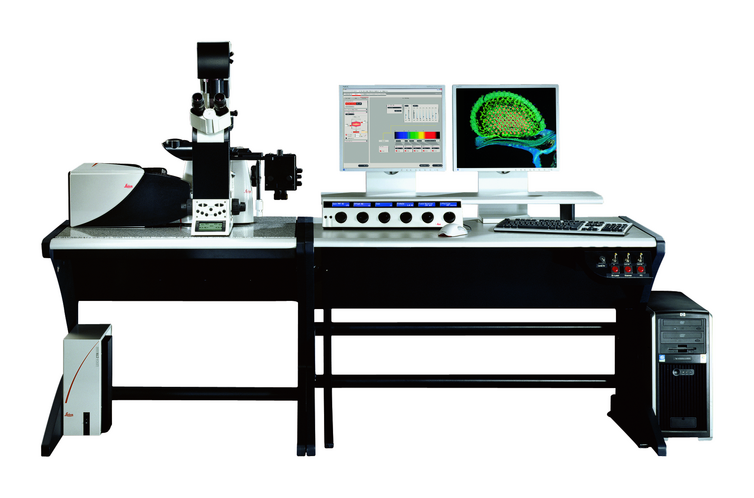
- Leica Confocal SP5 MP AFM
- Leica TIRF Thunder dSTORM
- Leica SP8 MP Dive
- Leica Confocal SP8-STED FLIM/FCS
- Leica TCS SP8 Digital Light Sheet Microscope
- Leica Confocal SP5 Blue
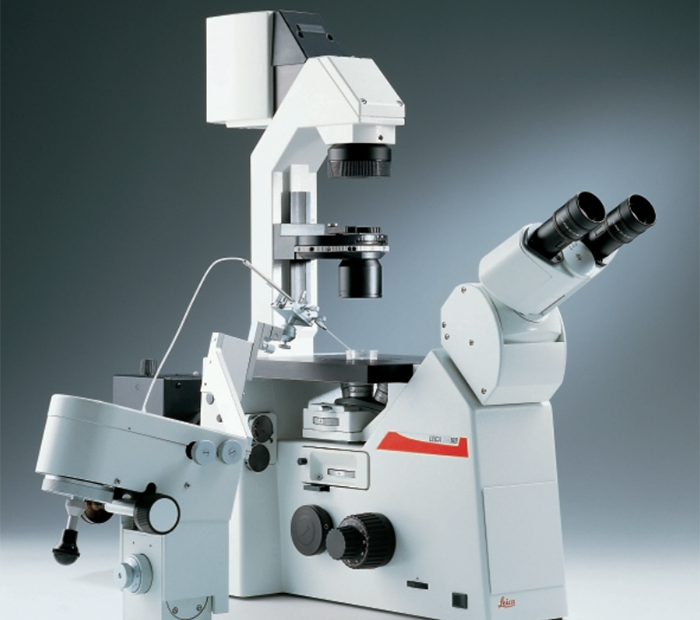
- Wide-Field CCD Microinjection Inverted Microscope
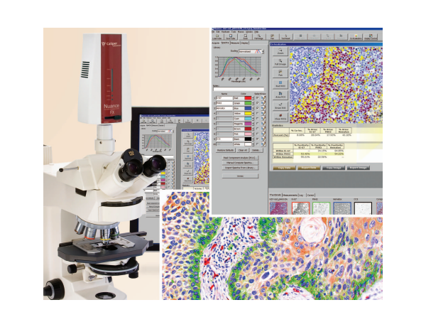
- Wide-Field NUANCE Microinjection Upright Microscope
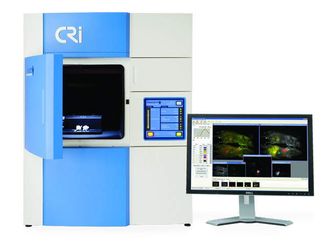
- Cambridge Research and Instrumentation Maestro 2
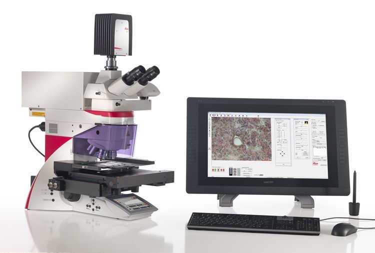
- Leica LMD7000 Laser Microsdissection System
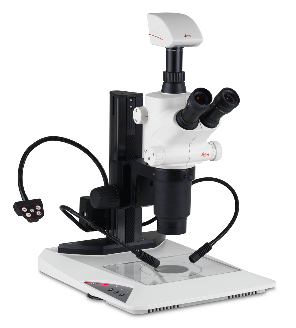
- Leica S8 APO Microscope
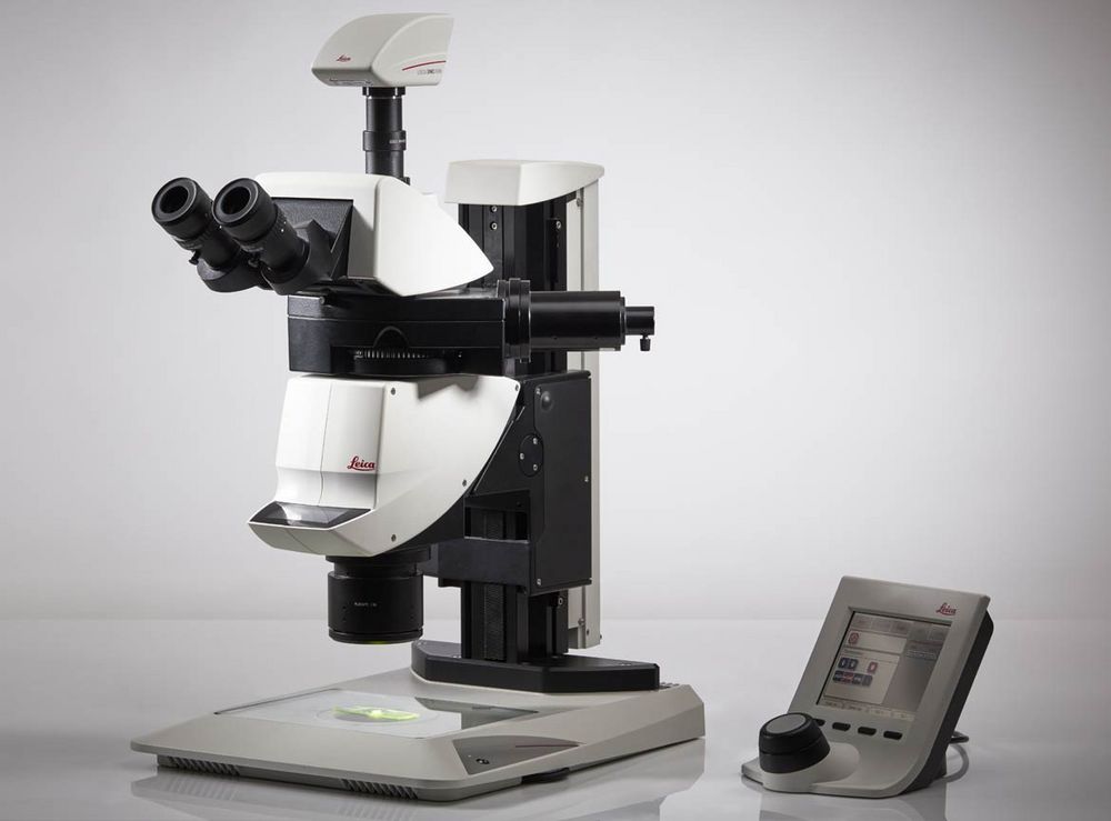
- Leica M205 FA Fluorescence Stereomicroscope
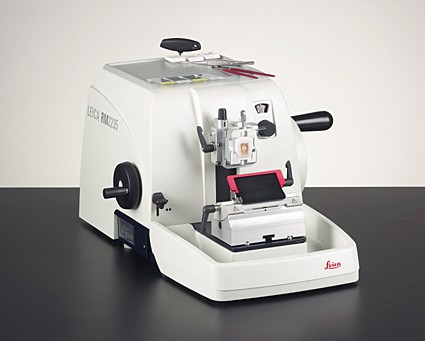
- Leica RM2235 Rotary Microtome
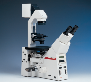
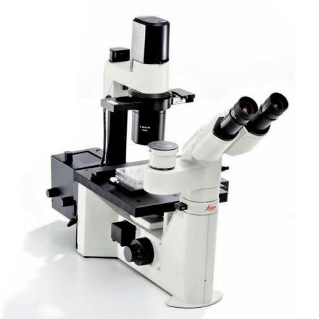
- Leica DM IL Inverted Microscope
- Leica LAS X Software Workstation
- Yokogaya confocal spinning disk
- AIVIA analysis workstation
- Sutter micropipette puller
- Tissue culture biosafety hood
Rates
| Instrument/Service | UC Affiliated User Rate | Non-UC Affiliated Academic/Non-Profit User Rate |
|---|---|---|
| Assisted Usage | $107 /hour | $147 |
| Confocal SP2 MP-FLIM | $44 /hour | $61 |
| Confocal SP5 Blue / MP-STED-AFM | $44 /hour | $61 |
| Confocal SP8 Light-Sheet / MP-DIVE-FLIM / SP8-STED/FLIM/FCS | $53 /hour | $74 |
| Consultation | $107 /hour | $147 |
| CRi Maestro 2 in-vivo imaging system | $34 /hour | $47 |
| Custom Projects: Lightsheet, Proof-of-Concept, SWIR imaging, Sample Analysis | $2,418 / project | $3,337 |
| Data/Image analysis/processing | $107 /hour | $147 |
| DMIL Microscope with SPOT Camera | $20 /hour | $27 |
| Equipment Loan – Tier 1 (Daily) | $53 /day | $74 |
| Equipment Loan – Tier 2 (Daily) | $108 /day | $149 |
| Equipment Loan – Tier 3 (Weekly) | $241 /week | $333 |
| Group Training | $160 /user /session | $221 |
| In Vivo Optix | $35 /hour | $48 |
| Leica DMI6000 confocal spinning disk | $44 /hour | $61 |
| Leica DMRXA Wide-field CCD IN SITU upright | $20 /hour | $28 |
| Leica LMD7000 | $44 /hour | $60 |
| Leica M205 FA fluorescence stereomicroscope | $20 /hour | $28 |
| Leica RM2235 Rotary Microtome | $19 /hour | $27 |
| Leica TIRF-STORM-Thunder | $55 /hour | $75 |
| Off Hour Rate – Confocal SP5 | $35 /hour | $48 |
| Off Hour Rate – Confocal SP8 | $44 /hour | $60 |
| Personalized one-on-one training (2 hours) | $213 /user/ session | $294 |
| SP8 Analysis Workstation | $20 /hour | $28 |
| Sutter Instruments micropipette puller | $20 /hour | $28 |
| Tissue Culture | $20 /hour | $28 |
| Wide-Field CCD Microinjection Inverted | $20 /hour | $28 |
| Wide-field Nuance microinjection upright | $20 /hour | $28 |