Brain Mapping Center
Overview
The Ahmanson-Lovelace Brain Mapping Center is the location where all of the scanning equipment used by the UCLA Brain Mapping Center is housed. The center includes a Siemens 3T Prisma scanner, a Siemens 3T Biograph mMR (PET/MR) scanner, a Cyclotron, a Transcranial Magnetic Stimulation Laboratory and an Electroencephalography Laboratory.
Leadership
Services
Imaging
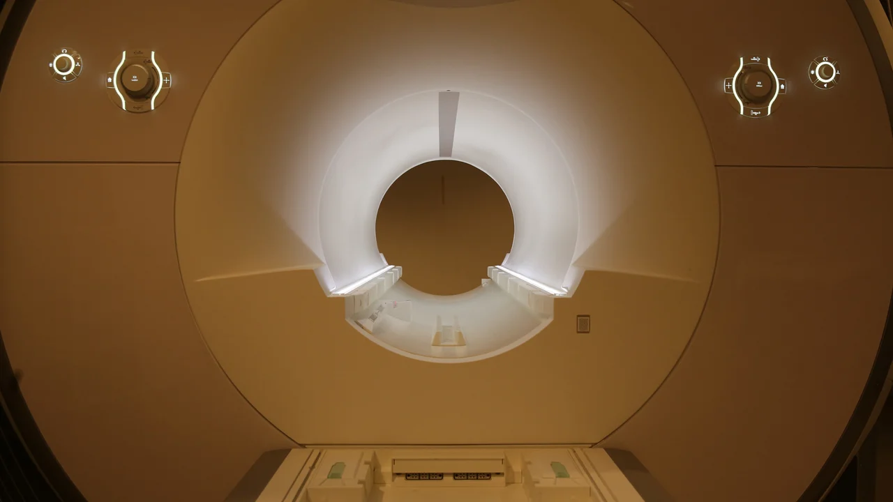
The Magnetic Resonance Imaging (MRI) laboratory, occupies approximately 4,500 sq. ft. of the Ahmanson-Lovelace Brain Mapping Center. This facility houses a Siemens Prisma 3.0 Tesla MRI Scanner.
The PET laboratory is in a suite specially designed for its use. It occupies a space of approximately 1668 square feet divided into a room for a cyclotron and its support equipment, a mMR scanner (PET/MR), a control room, uptake room/bathroom and a radiopharmaceutical lab.
Neuromodulation
The UCLA NeuroModulation Laboratory (NML) was founded in 1999 as part of the Ahmanson-Lovelace Brain Mapping Center as a novel brain mapping modality capable of mapping the brain function by using magnetic fields noninvasive brain stimulation. Since that time, a wide-variety of TMS protocols have been (and continue to be) developed to investigate brain neurophysiology, brain-behavior relationships, neuroplasticity, and potential treatment for some neurologic and psychiatric disorders.
In the UCLA NML, we have designed and implemented brain mapping protocols to study neurophysiology and brain-behavior relationships in projects involving motor control, visual perception, action-observation, language, impulsivity, spatial compatibility, often in collaboration with labs throughout UCLA. Among patient populations, we have and continue to study TMS effects in patients affected with Parkinson’s disease, parkinsonism, dystonia, epilepsy, stroke, mal de debarquement syndrome, migraine, and schizophrenia. Given the interest in using repetitive TMS for the potential treatment of neurologic and psychiatric disorders, in 2010, the TMS Lab collaborated in the design and implementation of the first major multicenter clinical trial of rTMS for Parkinson’s disease in North America (the MASTER-PD study).
Being in the ALBMC also provides a unique opportunity to combine TMS protocols with imaging (PET and fMRI studies) modalities since all are in close physical proximity to each other. Often, imaging studies are done first and then, using a frameless stereotaxy system, we can target pre-selected areas based on anatomic or functional coordinates within the brain. Alternatively, a modulatory train of TMS can be done and a rapid post-TMS imaging study can then be performed to assess for multimodal post-TMS effects.
What is TMS?
Transcranial magnetic stimulation (TMS) has been used since 1985 to study brain function by stimulating the brain painlessly and non-invasively. In TMS, an insulated coil is placed flat over the scalp. A brief current pulse is discharged through the coil. This results in a rapidly changing magnetic field that in turn induces an electrical current in the brain in the area directly underlying the coil. Each pulse is perceived as a touch or a tap to the scalp, resulting in an essentially painless procedure for stimulating the brain. For example, when the motor cortex is stimulated, there is an excitation of the underlying neurons which activate corticospinal neurons, resulting in muscle twitch on the muscles controlled by that region of the motor cortex.
TMS can be used over other parts of the brain to study a wide range of different behaviors, including visual perception, attention, language, and memory. TMS over specific locations in the frontal, temporal, parietal or occipital cortex can induce a transient perturbation in brain function, allowing us to create brain maps for each of these mental processes. Furthermore, the temporal resolution of TMS allows us to create brain maps that depict time as well as space. If a single pulse is delivered to particular brain regions at a variety of different time points during a mental process, we can develop maps that track the processing of information in the brain in both space and time. Different functional maps can be created at different times in the same person. This allows us to study the changes in the organization of the brains of people who are learning a new skill, and these data may help understanding plastic changes occuring in patients who have suffered injury or disease to the nervous system
In addition to the uses described above, TMS has also been applied to a wide variety of clinical research questions. These include localizing important movement and language areas for presurgical planning in patients with brain tumors or medically unresponsive epilepsy; understanding recovery after stroke or spinal cord injury; improving the diagnosis and treatment of movement or seizure disorders (e.g., Parkinson’s disease, epilepsy); and even treating patients with severe mental disorders such as depression or schizophrenia.
Data Analysis
(insert descriptive text)
Equipment
Siemens Prisma 3.0 Tesla MRI Scanner
The 3T Prisma scanner is a Magnetic Resonance Imaging system configured with up to 64 receiver channels comprised of up to 204 coil elements with up to 128 independent RF channels. It has has a 60 cm bore diameter and a 50x50x50 cm3 imaging field of view. It offers Diffusion Tensor Imaging (DTI) with up to 256 directions, Diffusion Spectrum Imaging (DSI) with up to 514 diffusion directions, remarkable flow sensitivity and time-resolved Angiography with XR 80/200, fully integrated funtional MRI/DTI evaluation, enhanced iPAT performance, powerful shimming with multinuclear spectroscopy, TimTX TrueShape and syngo ZOOMit with high resolution Diffusion Weighted Imaging (DWI) and long-term signal stability for demanding research sequences.
Siemens Biograph nMR Scanner (PET/MR)
The mMR scanner is in a suite specially designed for its use. It occupies a space of approximately 700 square feet divided into a room for a cyclotron and its support equipment, a mMR scanner, a control room, uptake room/bathroom and a radiopharmaceutical lab. Appropriate support rooms for patient waiting, reception and changing are available. Three-dimensional image acquisition and reconstruction are available with this device.
Adjacent to the mMR scanner suite resides a Siemens-CTI cyclotron. This device (RDS-111) is capable of producing a full spectrum of positron-emitting radioisotopes including: 15O, 13N, 18F and 11C. We currently produce sufficient quantities of oxygen-15 labeled water to perform cerebral PET activation studies. In addition, this includes an on-line continuous synthesis system capable of converting cyclotron-produced 15O into 15O-labeled water, carbon dioxide, carbon monoxide and oxygen gas for continuous or bolus inhalation or intravenous infusion (water) to the subjects. This system is fully automated and computer controlled thereby severely limiting or eliminating radiation exposure to personnel.
Imaging and TMS Rates
MRI Acquisition - 3T Scanner
- MRI = $700/hr
- Late Cancellation Fee = $140/hr (cancellation deadline is 24hrs prior to scan time)
- Prorated in 15min increments
mMR (PET/MR) Acquisition
mMR = $700/hr (same rate for PET, MRI only or both simultaneously)
- There is curently no charge for uptake time (please note this will be changing)
- 30min should be added to the scan acquisition time for setup/cleanup
- Late Cancellation Fee = $140/hr (cancellation deadline is 24hrs prior to scan time)
- Prorated in 15min increments
- Please note, these rates will be changing sometime in 2025 - if you planning a new budget please reach out to Mary Susselman (mwalker@mednet.ucla.edu) for more information.
- Radiopharmaceuticals can purchased separately from PETNET, the Biomedical cyclotron or other radiopharmacys
- FDG from PETNT costs: $155.10 for up to a 15mCi dose
Transcranial Magnetic Stimulation (TMS) Acquisition
- Data Acquisition = $300/hr
- Prorated in 15min increments