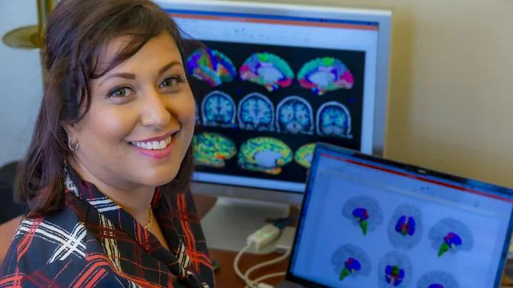
Neuroimaging Core
Goodman-Luskin Microbiome Center
Overview
We provide a wide range of state-of-the-art services to support researchers and clinicians interested in multimodal neuroimaging data acquisition, processing, and advanced analyses obtained from human studies related to existing data sets, ongoing studies, new studies, grant development, manuscript preparation, and data visualization, to name a few. Our analysis pipelines are able to handle processing from using various acquisition protocols.
Leadership

Contact
Email: MicrobiomeNeuroimagingCore@mednet.ucla.edu OR ArpanaChurch@mednet.ucla.edu
Website: Microbiome Neuroimaging Core
Services
- Consultation on data acquisition and experimental design
- Brain preprocessing of raw brain images
- Quality control
- Preprocessing
- Neuroimaging data will undergo quality control using our optimized and standardized Brain Imaging Data Structure (BIDS)-App, MRIQC and established protocols. BIDS-Apps have been developed to assess a wide range of quality control metrics, and implement a growing number of popular functional and structural MRI analyses
- Advanced neuroimaging methodologies including:
- Regional brain grey matter morphometry (structural magnetic resonance imaging)
- Functional (resting state and task-based fMRI)
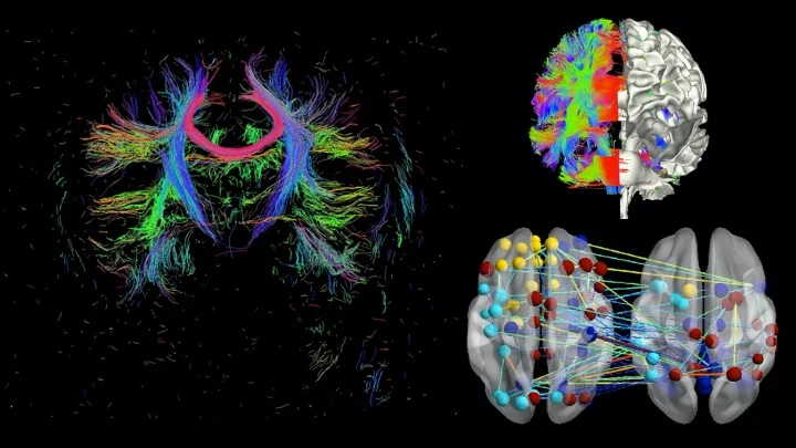
- Anatomical microstructural connectivity (diffusion spectrum imaging)
- Positron Emission Tomography
- Advanced multimodal data analyses and novel approaches for analyses of large multisite datasets such as:
- Network-based analysis (e.g., Graph theory-based association network analysis).
- Gradient analyses
- Myelinogenesis/Myelin Maps
- Neurometrics: integration of multiple analyses
- AI-powered tools to deconstruct datasets into detailed tissue maps, providing additional insights
- Sophisticated orchestra of data and insights. Our visualization tools transform intricate brain data into clear insights and build compelling visual narratives out of integrative, multi-omic studies.
- Data management and storage
- Assistance with interpretation of analysis results
- Research project development for manuscripts and grants
- Coordination with the other cores of the Microbiome Center (e.g., Data Core, Microbiome Core, and Bioinformatics and Biostatistics Core)
Equipment
The Neuroimaging Core (NIC) is housed in the UCLA Center for Health Sciences, 42-210.
Software required for state-of-the-art Magnetic Resonance Imaging (MRI) analysis techniques:
- Grey matter parcellation (Freesurfer), anatomical connectivity (diffusion tensor Imaging; Camino software), functional connectivity (resting state imaging; CONN software), graph theory based complex network analysis, connectivity gradient analysis, multivariate analysis.
- Established international imaging analysis programs that have been extended by custom scripting and internally developed algorithms. The library of available analysis programs provides all major mathematical, neuroimaging, and statistical packages, including AFNI, Matlab, R, SPM, FSL, Freesurfer, and many others.
- Utilize an integrated system of 112 CPU cores, 188 GB of RAM, and 80+ TB of cloud-hosted storage with automated nightly backups.
- Data transfer across servers via a gigabit storage area network (SAN).
- Additional storage resides on campus grid systems (grid.ucla.edu) and the UCLA cloud service (cloud.ucla.edu).
- No access to central repository data is available outside the UCLA firewalls.
- Collaboration via internet-accessible data stores occurs in a monitored, restricted network DMZ.